Eyelid Anatomy Explained: Layers, Muscles & Functions
Ever wondered what happens every time you blink?
Your eyelids are not simple skin flaps covering your eyes; they are layered, intricate, and important things that take care of your vision, hydrate them, and safeguard them. Your eyelid structure is critical in such activities as blinking, the production of tears, eye motion, and even facial expression.
In this complete guide to eyelid anatomy, I will carry you layer by layer, to demonstrate how your upper eyelids and lower eyelids are constructed, what they do, and why it is so important to pay attention to the design of your eyes afterward, whether you are a medical student, the patient on a table or someone with curiosity, wishing to know how your body works.
What Is an Eyelid?
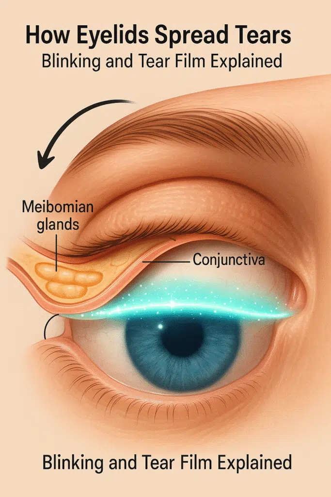
The cornea is a shielding and elastic film that covers the surface of your eye and is instrumental in ensuring that it is clean, safe, and healthy. Whenever you blink, your eyelids, such as your lower and upper eyelids, assist in covering the tears of your eye, getting rid of dust or other particles, and eliminating harmful light. Without them, your eyes will dry up, become itchy, and run the risk of compromised conditions.
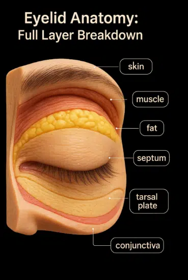
The eyelid consists of skin, muscles, fat, glands, and a layer of tissue lining found on the inside called the conjunctiva. All the components in these are integrated to nourish your eyes and promote sight.

Layers
There are three eyelids, top and bottom, to each eye:
- The upper lid moves more and aids in the blink of your eye.
- Less mobile, yet important, is the lower eyelid.
- The external appearance of the eyelid.
Function of Eyelids
The following are the functions of the Eyelids:
- They guard the eye against foreign objects and trauma as well as unnecessary light.
- They aid in the distribution of tear film, and this makes the cornea moist and clean.
- They endorse facial expression, particularly by blinks and movement.
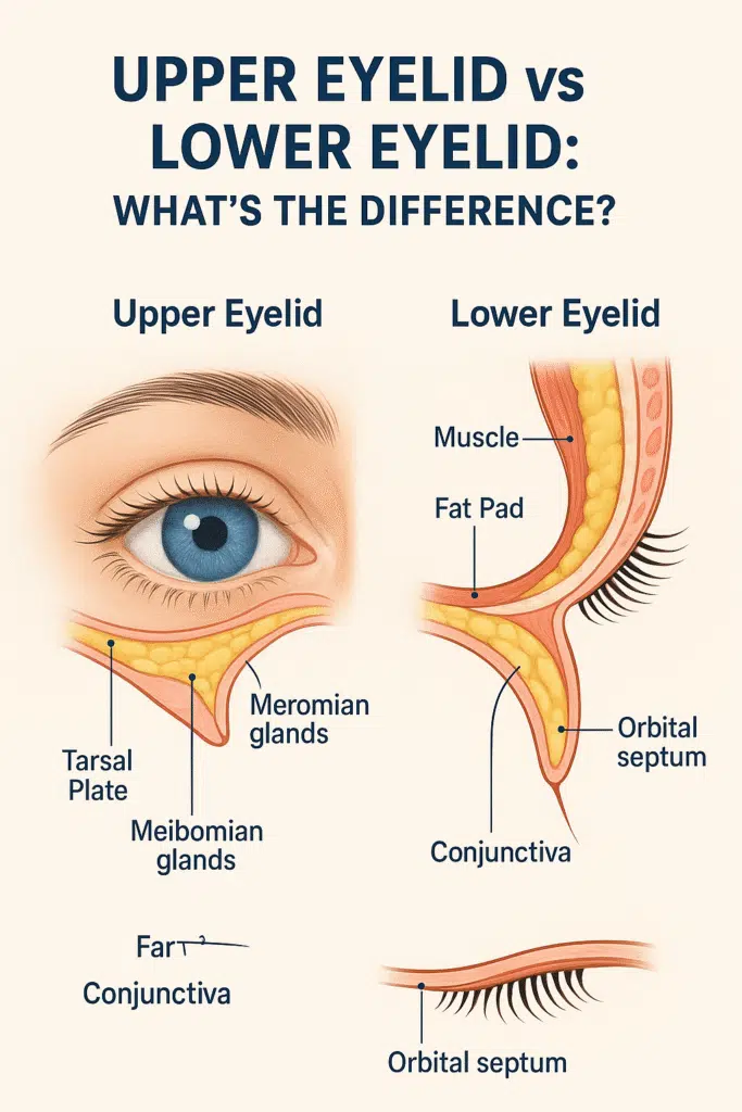
Upper Eyelid Anatomy
The upper eyelid is probably the least important part of your face, but it is composed of multiple layers that all provide functions that help to protect your eye, keep it properly moistened, and help you blink quite smoothly. This is more mobile and complicated than the lower eyelid, and there is a purpose to each of the parts.
Skin
- Your upper eyelid skin is the thinnest on your entire body.
- It is too soft and supple, and it is loosely attached to those layers beneath it.
- .Such a loose grip enables them to move freely with your facial expression, and as you blink your eyes.
- .It is that mobility that provides the upper eyelid with the capacity of smooth folding.
Subcutaneous Areolar Tissue
- Under the skin, a very thin layer of loose connective tissue exists.
- This is the region that contains very little fat, which is why the upper eyelid appears to be so delicate.
- It assists the skin in sliding against underlying muscles easily.
Muscles of the Upper Eyelid
Your eyelid is made of strong muscles and is capable of making your eyes blink and protecting them. This is what they are doing:
Orbicularis Oculi
- It is a round muscle that helps to shut/squeeze your eyelids when you blink, squint your eyes, or close them.
- There are three major sections in it:
- Orbital part – it assists you to close your eyes tightly.
- Preseptal part –it contributes to the normal blinking.
- Pretarsal part –performs gentle and unconscious blinking to allow the spread of tears.
Levator Palpebrae Superioris
- This is the muscle that opens the eyelid of the upper eye.
- It begins inside the center of your eye at the back of the socket and grows out.
- It terminates in a tendon named the levator aponeurosis, which:
- It attaches to the tarsal plate (which is the rigid support of an eyelid).
- Insert in the skin of the eyelids and create the upper eyelid crease that you observe.
Müller’s Muscle
- There is a thin smooth muscle beneath the levator aponeurosis
- It provides you with a little bit of extra elevation to your upper eyelid.
- It is under the control of the sympathetic nervous system (stress responding).
- When it fails to work, your upper eyelid might droop down a little bit and this is known as ptosis.
Orbital Septum
- It is a fibrous sheet that runs between the eyelid and the further reaches of your eye socket.
- It extends from the bony outline of the orbit to the tarsal plate.
- It makes a shield, keeps the orbital fat and muscles well held in, and guards your eye against outside infection and injury.
Pre-Aponeurotic Fat Pads
- These fat pads occupy the immediate position behind the orbital septum.
- In the upper eyelid, two compartments are found:
- Medial fat pad
- Abdominal fat pad
- They act as cushions to your eye, secure it against pressure, and keep the eyelid with its smooth contour.
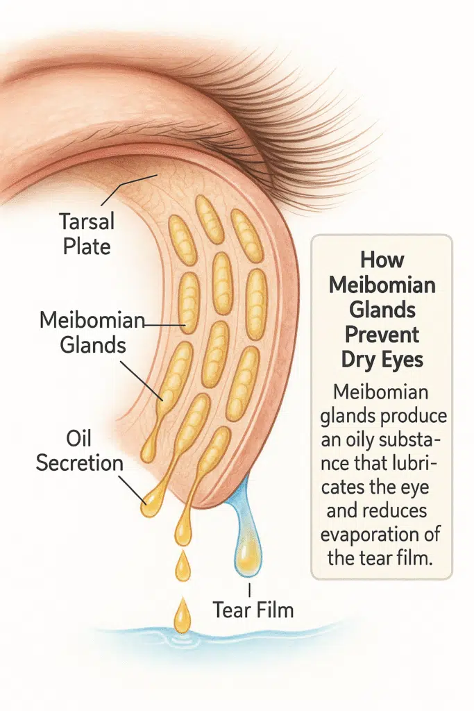
Tarsal Plate
- It is a strong, hard bit of connective tissue, sort of your eyelid skeleton.
- In the upper eyelid, it measures about 10-12 mm.
- It provides your eyelid with a stable shape and maintains it firm
- The Meibomian glands found within the tarsal plate are said to play an important role in the stability of tears:
- They make up the oily component of your tear film and prevent the drying of your eyes.
- In the absence of this layer of oil, your tears would dissipate too quickly.
Conjunctiva
- This is the shiny, smooth lining on the surface of your eyelid.
It is known as the palpebral conjunctiva and reflects on the eyeball, becoming the bulbar conjunctiva.- It keeps your eyelid and eye surface moistened, healthy, and safe.
- It is also a shield against dust, bacteria, and allergens.
Read Also: LASIK Eye Surgery Risks & Side Effects You Must Know
Why Knowing Upper Eyelid Anatomy Matters
When you understand the structure of your upper eyelid, you start to see just how important it is. Every blink you make depends on the precise coordination of muscles, fat pads, glands, and connective tissue — all working behind the scenes
This is what I have written to you; whether it is to study a medical career, planning to have an eyelid procedure, or purely out of curiosity to know more about your body, I have written to you so that you may be able to see the importance, and usage of something you use every second of every day.
Lower Eyelid Anatomy
The lower eyelid is very similar to the upper lid but there are significant differences in terms of the height as a whole, muscle placement, and tissues employed to support the eyelid.
Skin and Subcutaneous Tissue
- The skin, like the upper lid, is thin and movable.
- Flexibility and mobility are possible due to loose areolar connective tissue.
Orbicularis Oculi
- The same as in the upper eyelid.
- Important in facial expression, tear pumping, and eye closure.
Orbital Septum
- Prolonged on the orbital rim to the inferior tarsal plate.
- Serves as a shield to the orbital structures.
Tarsal Plate
- Not so long as those of the upper lid, approximately 4-5 mm long.
- Meibomian glands such as on the upper lid are housed.
Conjunctiva
- Constitutes the inner lining of the lower lid.
- Keeps in touch with the bulbar conjunctiva at the inferior fornix.
Pre-Aponeurotic Fat Pads
- The pre-Aponeurotic Fats Pads include:
- Medial
- Central
- Lateral
- The lower eyelid contains three fat pads as opposed to the upper eyelid, which has one fat pad.
Lower Eyelid Retractors
- Similar to the upper lid levator and Mollé muscles.
- The projections of the inferior rectus muscle sheath.
- Assistance in depression of the lower eyelid in downward gaze.
External Eyelid Anatomy
The outer appearance of your eyelids determines the visible outline of the eyes and also serves as a part of functionality and expressivity.
Eyelid Margins
- These are the margins of eyelids, and they possess special anatomical points:
- Eyelashes (Cilia): Trap dust and debris.
- Meibomian Gland Orifices: ducts of secretion of tear lipids.
- Gray Line: A dark line between the eyelid structures of the front and back. It is a very important surgical landmark.
Eyelashes (Cilia)
- Appearing in rows of three on the margin
- Aid in providing guard against the effects of air particles and light on the cornea..
- There is a sebaceous gland (Gland of Zeis) that is attached to each Lash.
Eyelid Crease
- Presented only in the upper lid.
- The form of an augmentation made by insertion of the levator aponeurosis into the skin.
- This crease determines the shape of the visible eyelid, and it differs according to ethnicity and age.
Lacrimal Puncta
- Small holes on the medial margin of each of the lower and upper eyelids drain tears.
- Perch upon tiny heights referred to as lacrimal papillae.
- Into the canalicular system then into the nasolacrimal duct.
Medial Canthus
- The inside of the eye.
- Contains:
- Lacrimal Caruncle: It is a pink fleshy bump that bears sebaceous glands.
- Plica Semilunaris is a relic of the third eyelid, a conjunctival groove in some animals.
Lateral Canthus
- The peripheral dark one of the eyes.
- Brings the upper eyelid and the lower eyelid together at the lateral orbital edge.
- The skin may be anchored to bones through the lateral canthal tendon.
Palpebral Fissure
- The interval between the open eyes.
- It has different shapes and sizes of people, and also different faces.
- Typically, around 8–10 mm vertically and 28–30 mm horizontally.
Eyebrows
- They are not part of the eyelid but have a protective function.
- It is placed on the supraorbital ridge.
- Assist in preventing sweat and moisture on your eyes.
- In addition, contributes to facial expressions and non-verbal communication.
FAQs
What is the function of the eyelids?
Eyelids save your eyes against dust, dry air, and light, and also help to spread the tears in your eyes to make your eyes moist and healthy.
Which muscles move the eyelids?
The orbicularis oculi causes the eyelids to close. The superior levator palpebrae and the Müller muscle are the ones that elevate the upper eyelid.
Why is the upper eyelid more mobile?
It is more vigorous than the lower eyelid since it has more powerful muscles to lift, among them levator palpebrae.
What do Meibomian glands do?
They secrete the oily film of your tears, which ensures that your tears do not dry up and stabilizes them.
What is the tarsal plate?
A thickened substance that provides the shape of the eyelid and fixes the Meibomian glands.
Read Also: Can Myopia Get Better with Age? The Truth Revealed!
Final Words
Your eyelids are tiny, though their structure is not at all straightforward. Just as healthy skin, fragile as it is, is on the surface of your eyelid, there are deeper muscles, fat cushions, and glands that take care of your eyes and ensure their healthy sight and comfort in day-to-day life.
Before eyelid surgery or just out of simple curiosity as to how your body functions, one could ask how the upper and lower eyelids are structured.